All published articles of this journal are available on ScienceDirect.

Alternating Hemiplegia of Childhood and Developmental Delay: A Longitudinal Pediatric Neurogenetic Case Report
 iD
Authors Info & Affiliations
Abstract
Background
Alternating hemiplegia of childhood (AHC) is a rare neurodevelopmental disorder most commonly caused by pathogenic variants in ATP1A3. It is characterized by recurrent episodes of alternating hemiplegia, paroxysmal neurological manifestations, developmental delay, and epilepsy. Access to molecular diagnosis remains limited in many low- and middle-income countries, contributing to delayed recognition and management.
Case Presentation
This case report describes a female patient with paroxysmal alternating hemiplegic episodes, developmental delay, and epilepsy. Hemiplegic events were characterized by alternating laterality and remission during sleep at 2 years and 10 months of age. Neuroimaging was largely unremarkable, including a structurally normal brain CT at 14 years of age, without evidence of vascular or progressive structural pathology. Genetic testing identified a heterozygous pathogenic variant in ATP1A3 (c.2839G>A; p.Gly947Arg), confirming the diagnosis of ATP1A3-related AHC. An additional CDKL5 c.2156C>T variant of uncertain significance was detected but was not clinically contributory based on phenotype correlation. Multidisciplinary follow-up showed partial response of hemiplegic episodes to flunarizine and preserved cardiac structure and function.
Conclusion
This case reinforces the clinical and genetic heterogeneity of ATP1A3-related alternating hemiplegia of childhood and highlights the importance of molecular confirmation for accurate diagnosis and management. In resource-limited settings, delayed access to genomic testing may prolong the diagnostic odyssey. Strengthening neurogenetic diagnostic capacity in Latin America is essential to improve early recognition, guide precision-based treatment strategies, and reduce healthcare inequities for patients with rare neurodevelopmental disorders.
1. INTRODUCTION
Alternating Hemiplegia of Childhood (AHC) is a rare neurodevelopmental disorder characterized by recurrent hemiplegic episodes, paroxysmal neurological manifestations, and progressive neurodevelopmental impairment [1, 2]. The disorder typically presents within the first six months of life and is frequently misdiagnosed during early childhood due to its clinical heterogeneity and overlap with epilepsy, migraine, and metabolic encephalopathies [3, 4]. Hemiplegic attacks recur throughout the patient’s lifespan; however, their frequency and duration often decrease with advancing age. In addition to motor manifestations, many patients exhibit distinctive facial features, including elongated face, thin eyebrows, and hypertelorism (Fig. 1a) [5]. Gastrointestinal involvement is also common, presenting as vomiting, dysphagia, anorexia-related weight loss, nausea, and drooling, with additional minor symptoms such as constipation, diarrhea, and abdominal pain [6, 7]. Persistent gastrointestinal dysfunction may generate a vicious cycle that exacerbates clinical severity and increases susceptibility to infections. The diagnostic framework for AHC relies on clinical criteria, exclusion of differential diagnoses, and molecular confirmation, most commonly involving pathogenic variants in ATP1A3 (Fig. 1b). Despite advances in genetic testing, delayed or incorrect diagnosis remains frequent, particularly in low- and middle-income countries. Here, we report an adolescent female from Ecuador with genetically confirmed AHC, initially misdiagnosed, highlighting the importance of early recognition and molecular confirmation [8].
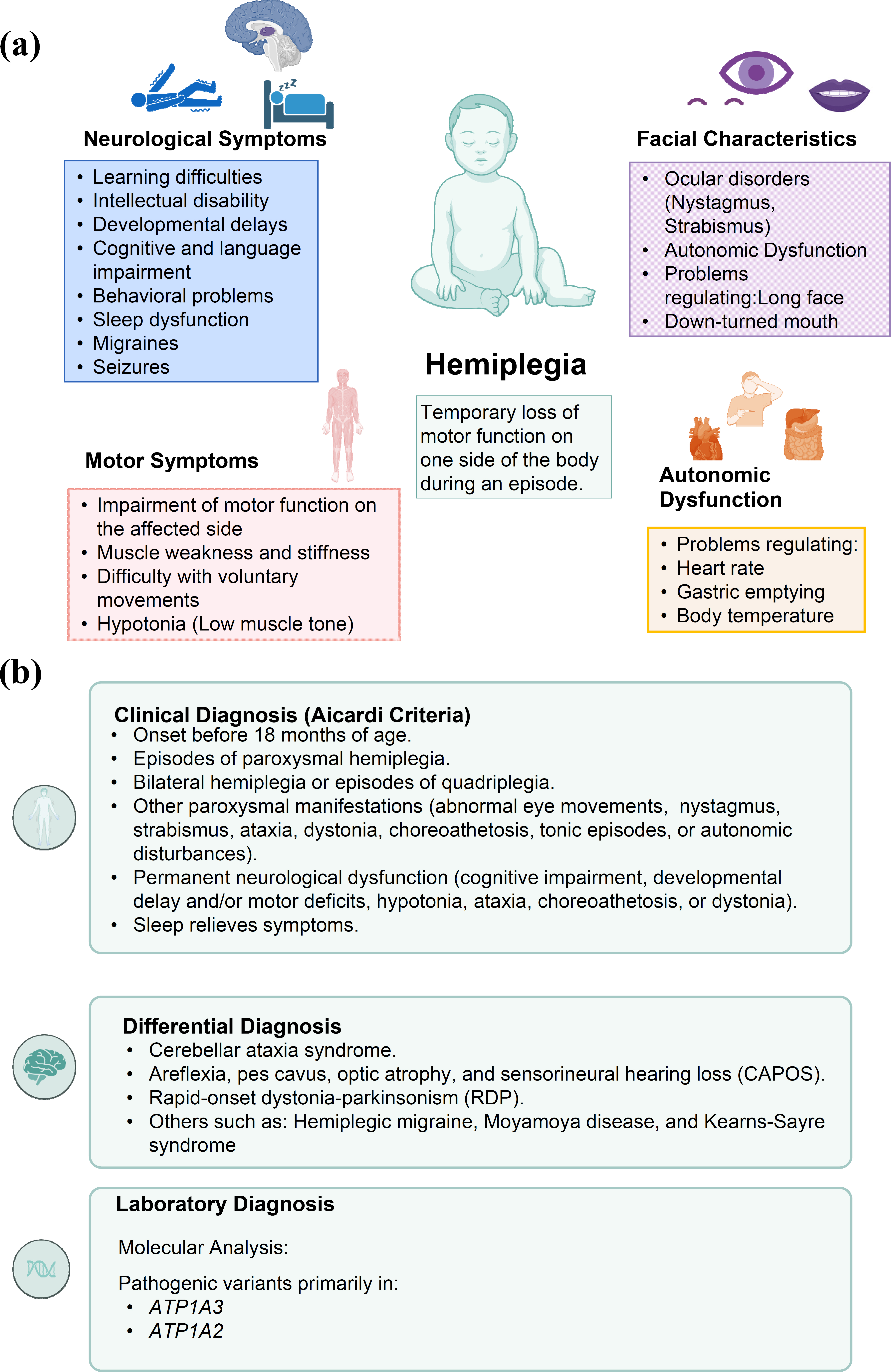
Clinical features and diagnostic framework of alternating Hemiplegia of Childhood (AHC). (a) Color-coded clinical domains: neurological manifestations are shown in blue, motor manifestations in pink, facial and ocular characteristics in purple, autonomic dysfunction in yellow, and the central definition of hemiplegia in green. (b) Diagnostic workflow beginning with the Aicardi clinical criteria, followed by exclusion of alternative neurological disorders and molecular confirmation. The upper icon represents clinical assessment, the lower-left brain icon represents neurological and neuroimaging evaluation of differential diagnoses, and the lower-right DNA icon represents genetic testing. Abbreviations: AHC, alternating hemiplegia of childhood; ATPase Na+/K+ transporting subunit alpha 2; ATP1A3, ATPase Na+/K+ transporting subunit alpha 3 [9].
2. CASE PRESENTATION
The patient is a 17-year-old female adolescent from Ecuador, a middle-income country with limited access to pediatric neurogenetic services. She presents age-appropriate physical growth but marked cognitive, intellectual, and social developmental delay with expressive language impairment. A detailed clinical timeline is provided in Table 1.
| Age | Clinical Events | Investigations | Treatment | Outcome |
|---|---|---|---|---|
| 2 years, 10 months | Onset of alternating hemiplegic episodes | Clinical evaluation | Symptomatic management | Recurrent, self-limited episodes |
| 3 years | Prolonged hypotonic/hemiplegic episodes with gaze deviation and nystagmus; developmental delay | Neurological follow-up, multiple Exams | Supportive care; temporary low-protein diet, L-carnitine, vitamin B12, coenzyme Q10, and lactulose | Metabolic, infectious, and vascular causes were not supported; episodic course persisted |
| 4 years | Clinical diagnosis of AHC | Clinical criteria assessment | Flunarizine initiated | Partial reduction in attacks |
| Adolescence | Seizure onset | Brain MRI; EEG | Levetiracetam | Seizure control achieved |
| 11 years | Molecular confirmation | ATP1A3 p.Gly947Arg (heterozygous, pathogenic); CDKL5 VUS | Continued therapy | Genetic diagnosis established |
| 14 years | First generalized tonic-clonic seizure | Hospital evaluation; brain CT | Levetiracetam initiated | Sustained seizure control |
| Latest follow-up | Intermittent hemiplegia; stable epilepsy | Periodic follow-up | Ongoing flunarizine and levetiracetam | Stable condition |
No family history of alternating hemiplegia, epilepsy, neurodevelopmental disorders, movement disorders, or other hereditary neurological diseases was reported by the patient’s caregivers.
She was born at term (39 weeks) via cesarean section with immediate crying and normal Apgar scores. Neonatal history was significant for maternal–fetal Rh incompatibility (patient A Rh negative). Psychomotor development was delayed: head control at 3 months, independent sitting at 8 months, crawling at 11 months, and autonomous ambulation at 22 months. The first hemiplegic episode occurred at 2 years and 10 months. Events consisted of sudden unilateral weakness with alternating laterality, predominantly affecting upper limbs, associated with axial hypotonia, transient facial asymmetry, dysarthria, pallor, diaphoresis, irritability, and reduced responsiveness. Episodes lasted 2–12 hours and were triggered by fatigue, emotional stress, heat exposure, and prolonged water immersion. Symptoms consistently resolved with sleep. At 3 years, she was hospitalized during a prolonged episode characterized by generalized hypotonia, horizontal nystagmus, gaze deviation, and sequential right- and left-sided weakness. During that admission, serum glucose was 78.67 mg/dL and cerebrospinal fluid was clear, with glucose 55.9 mg/dL, total protein 0.10 g/dL, no visible cells or bacteria on staining, and negative bacterial culture. A transient hyperammonemia prompted metabolic evaluation; however, plasma amino acids, urinary organic acids, newborn/metabolic screening, and the acylcarnitine profile did not support an inherited metabolic disorder. A prolonged sleep EEG at 3 years showed a normal background without epileptiform activity. Serial ancillary investigations helped exclude structural, metabolic, neuromuscular, sensory, and cardiac mimics. Angio-CT at 3 years showed no intracranial vascular malformation, a non-contrast brain MRI at 3 years was normal, and MR angiography at 3 years and 7 months demonstrated an intact circle of Willis and no diffusion abnormalities, with hypoplasia of the left lateral sinus interpreted as a normal variant. At 4 years, brain MRI revealed mild left temporal polar cortical atrophy, with preserved gray–white differentiation, normal hippocampi, no diffusion restriction, normal spectroscopy, and no arterial abnormalities. Neurophysiological studies showed normal motor and sensory nerve conduction, normal EMG, and normal visual evoked potentials. Audiometry was normal. Holter monitoring demonstrated sinus rhythm with rare isolated monomorphic ventricular ectopy and no pauses, and echocardiography showed preserved ventricular function with only mild tricuspid and pulmonary regurgitation. A clinical diagnosis of AHC was established based on the neurological phenotype according to ECC. The background was within normal limits for age, with no interhemispheric asymmetry, pathological slowing, or paroxysmal discharges. Importantly, a left upper-limb hemiplegic episode occurred without an electrographic correlate, supporting the non-ictal nature of the event (Fig. 2). Flunarizine was initiated (5 mg/day), resulting in a partial reduction in attack frequency and severity.
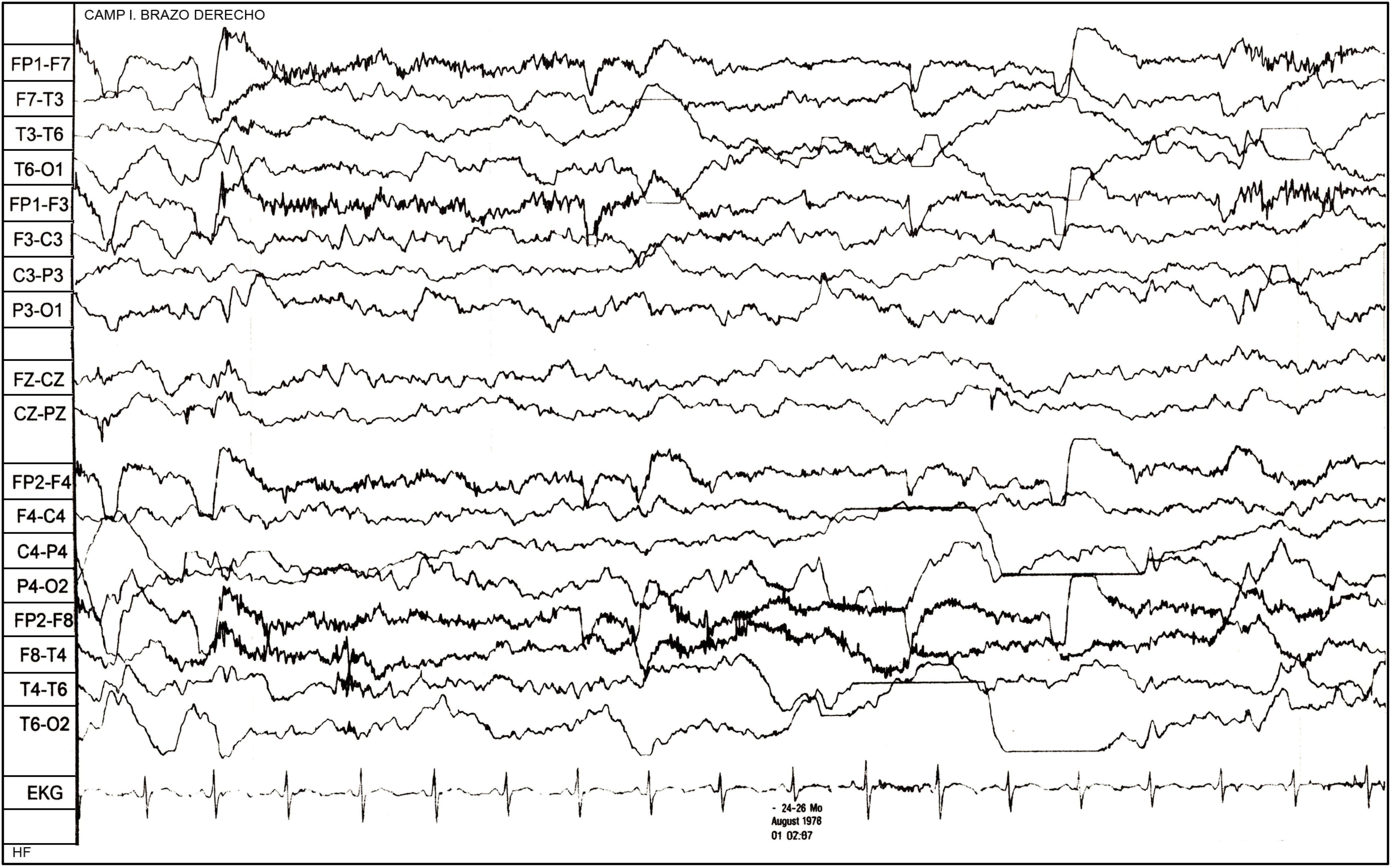
Representative electroencephalographic tracing obtained during the evaluation of the generalized tonic-clonic seizure, at 4 years.
Brain MRI revealed mild left temporal polar cortical atrophy, with preserved gray–white differentiation, normal hippocampi, and no diffusion restriction. No vascular abnormalities were observed. Neurophysiological studies showed normal motor and sensory nerve conduction, normal F-wave latencies, absent bilateral tibial H-reflexes, and normal needle electromyography. There was no evidence of peripheral neuropathy. Transthoracic echocardiography demonstrated normal cardiac structure and preserved systolic function (LVEF 77%), without cardiomyopathy or pulmonary hypertension.
Between ages 4 and 11, hemiplegic crises persisted with reduced intensity. At 11 years, prolonged syncopal episodes accompanied by mood instability prompted dose escalation of flunarizine and addition of clonazepam.
Genetic testing was performed using Illumina sequencing-by-synthesis technology with the Blueprint Genetics Comprehensive Epilepsy Panel rather than whole-exome sequencing. According to the laboratory quality report, the nuclear-genome component covered 474 genes and 7,147 exons/regions, comprising 1,398,564 target bases; 1,396,449 bases were sequenced. The mitochondrial-genome component covered 37 genes and 15,358 target bases; all 15,358 bases were sequenced. The panel included the clinically relevant AHC and differential-diagnosis genes ATP1A3, ATP1A2, SLC2A1, CACNA1A, PRRT2, SLC1A3, ADCY5, TANGO2 and TBC1D24; no pathogenic or likely pathogenic variants were detected in the alternative genes.
The assay identified a heterozygous ATP1A3 variant (NM_152296.4:c.2839G>A; p.Gly947Arg), classified as pathogenic by the external laboratory under an ACMG/AMP-based interpretation framework and concordantly classified as pathogenic in ClinVar (Table 2) [9, 10]. An additional heterozygous CDKL5 variant (NM_003159:c.2156C>T; p.Pro719Leu) was reported as a variant of uncertain significance and was not considered explanatory after phenotype correlation (Table 2). Testing was performed in the proband only. Parental segregation analysis was not performed because of limited access to testing and financial constraints; therefore, de novo occurrence could not be confirmed. Nevertheless, the pathogenic ATP1A3 p.Gly947Arg variant is presumed to be de novo, in line with the inheritance pattern described in most ATP1A3-related alternating hemiplegia of childhood cases [10].
| Gene | Variant (cDNA) | Genomic Location | Protein Change | dbSNP ID | Zygosity |
Variant Classification ACMG/ ClinVar |
Clinical Correlation |
|---|---|---|---|---|---|---|---|
| ATP1A3 | c.2839G>A | chr19:41967744 (GRCh38.p14) | p.Gly947Arg | rs398122887 | Heterozygous | Pathogenic | Yes |
| CDKL5 | c.2156C>T | chrX:18631275 | p.Pro719Leu | — | Heterozygous | Variant of uncertain significance (VUS) |
No |
At 14 years, the patient experienced a severe generalized seizure requiring hospitalization. Levetiracetam was initiated, achieving sustained seizure control over two years. Preventive measures consisted of structured rest and the avoidance of physical exertion, intense emotions (such as excessive joy), and prolonged water exposure. Also, a non-contrast Computed Tomography (CT) scan of the brain was performed as part of clinical follow-up. The brain CT showed no pathological hyperdensities or hypodensities. No intracranial hemorrhagic collections or space-occupying lesions were identified. The sellar and parasellar regions were preserved, without structural abnormalities. Midline structures were not displaced. There was appropriate gray–white matter differentiation. The cortical sulci and Sylvian fissures were preserved. The ventricular system and basal cisterns were within normal limits. The posterior fossa, fourth ventricle, and cerebellum showed no abnormalities. No relevant findings were identified on bone window imaging. Overall, the brain CT was structurally normal, with no evidence of acute or chronic vascular events, intracranial hemorrhage, mass lesions, or significant cortical atrophy. These findings are consistent with the pathophysiology of alternating hemiplegia of childhood, in which hemiplegic episodes are functional and paroxysmal in nature, secondary to neuronal dysfunction related to impairment of the Na+/K+-ATPase encoded by the ATP1A3 gene, rather than structural ischemic processes. The absence of structural abnormalities further supports that the clinical episodes do not correspond to cerebrovascular events or progressive structural brain pathology. Corrective surgery for genu valgum led to a significant improvement in gait stability. At last follow-up, the patient continues to experience intermittent hemiplegic episodes with partial response to flunarizine, while epilepsy remains well controlled. Flunarizine was used for prophylaxis of hemiplegic episodes and increased to 10 mg/day. Clonazepam was added after prolonged syncopal episodes and irritability. Levetiracetam (500mg/12h) was initiated after a generalized tonic–clonic seizure at 14 years, achieving sustained seizure control. Treatment adherence and tolerability were assessed through caregiver report during clinical follow-up and review of the available medical records. No standardized adherence scale was applied. According to caregiver-reported information and clinical documentation, the patient continued pharmacological management with flunarizine and levetiracetam, with partial reduction of hemiplegic episodes and sustained seizure control. No treatment discontinuation due to intolerance was documented.
No adverse or unanticipated treatment-related events were documented in the available clinical records. The seizures were considered part of the disease course rather than a treatment-related adverse event.
3. DISCUSSION
Population-based epidemiological data for AHC in Ecuador and Latin America remain unavailable. International estimates vary according to the denominator and ascertainment method. A nationwide Danish pediatric study identified 10 patients aged 16 years or younger and estimated a prevalence of approximately 1 per 100,000 children [11]. Differences in ascertainment may reflect clinical heterogeneity, historical underrecognition, unequal access to molecular confirmation, and underdiagnosis. Consequently, isolated Latin American case reports cannot be used to calculate regional incidence or prevalence [7, 12, 13]. In Ecuador, barriers to genetic testing, funding, and research on rare and undiagnosed diseases have been reported [14]. The relevance of this case does not rest solely on the identification of p.Gly947Arg, as this is a recurrent pathogenic ATP1A3 variant that has already been reported in Latin American patients. Nevertheless, to the best of our knowledge, the present study represents the first full-length longitudinal report of genetically confirmed p.Gly947Arg-related alternating hemiplegia of childhood in an Ecuadorian patient.
The distinctive contribution of this case lies in the characterization of a comparatively mild and slowly evolving phenotype, defined by the unusually late onset of alternating hemiplegic episodes at 34 months of age, epilepsy emerging during adolescence with sustained seizure control under levetiracetam, and only mild focal temporal atrophy after a prolonged disease course.
ATP1A3 encodes the neuronal Na+/K+-ATPase α3 subunit, which is particularly important for restoring transmembrane sodium and potassium gradients after sustained or high-frequency neuronal firing. Gly947 is located in the ninth transmembrane domain near the cation-binding region; replacement of a small glycine residue by a bulky, positively charged arginine is expected to disturb the conformational transitions required for efficient ion transport [10, 15]. Reduced α3-pump reserve provides a mechanistic framework for the patient's paroxysmal phenotype: heat, physical exertion, emotional stress, and prolonged water immersion increase neuronal firing and metabolic demand, thereby exceeding the capacity of the mutant pump to restore ionic gradients. Conversely, sleep reduces synaptic activity and energy demand, allowing the remaining pump reserve and other Na+/K+-ATPase isoforms to re-establish membrane homeostasis. This demand-reserve model plausibly explains both the reproducible triggers and the characteristic resolution of attacks during sleep, although it was not directly tested in this patient.
The comparatively mild disease is consistent with established genotype-phenotype correlations. Large cohorts have associated p.Gly947Arg with more favorable motor and intellectual outcomes than p.Glu815Lys, which is linked to the most severe AHC phenotype [16, 17]. Importantly, this difference cannot be explained simply by normal residual pump activity. In functional studies, p.Asp801Asn, p.Gly947Arg, and p.Glu815Lys all markedly reduced forward pump cycling and exerted dominant-negative effects; however, proton transport was selectively and substantially impaired by p.Glu815Lys, but preserved with p.Asp801Asn and p.Gly947Arg [18]. Other experimental data indicate that protein misfolding, endoplasmic-reticulum retention, unfolded-protein responses, and competition between α-subunit isoforms can further modify disease severity [9]. Thus, p.Gly947Arg may produce a narrower functional defect, with sufficient effective pump reserve under basal conditions but episodic failure under stress, whereas p.Glu815Lys adds disturbances that promote more persistent neuronal dysfunction. This interpretation remains inferential because patient-derived functional studies were not performed.
Longitudinal EEG findings help distinguish the paroxysmal hemiplegic events from comorbid epilepsy. EEG at 4 years captured left upper-limb hemiplegia without an evolving electrographic correlate and showed a normal age-appropriate background. This observation supports the concept that AHC hemiplegic attacks reflect transient dysfunction of neuronal ion homeostasis rather than focal ictal activity, while acknowledging that interictal epileptiform abnormalities and epilepsy may coexist independently. The generalized tonic-clonic seizure at 14 years should therefore be regarded as later comorbid epilepsy, emerging approximately a decade after the early hemiplegic phenotype, rather than as continuation of an unrecognized ictal disorder.
The late appearance of epilepsy also requires a cautious mechanistic interpretation. Epilepsy in AHC is heterogeneous and may evolve independently from hemiplegic attacks; moreover, epileptiform EEG abnormalities can emerge well after the first clinical seizures [19]. In this patient, the first generalized tonic-clonic seizure at 14 years suggests that cortical networks remained below the seizure threshold throughout early childhood despite recurrent paroxysmal motor events. A plausible explanation is that the partial functional reserve associated with p.Gly947Arg limited early network hyperexcitability, while cumulative ionic and metabolic stress, brain maturation, and patient-specific genetic or environmental modifiers eventually lowered the seizure threshold. Nevertheless, late epilepsy should not be considered a variant-specific feature of p.Gly947Arg, because substantial interindividual variability exists even among patients carrying the same recurrent variant. The absence of early refractory seizures or recurrent status epilepticus and the sustained response to levetiracetam support a lower epileptogenic burden than that observed in severe ATP1A3-related developmental and epileptic encephalopathy.
The neuroimaging findings are likewise compatible with a relatively attenuated course. Brain MRI in AHC is often normal, whereas progressive frontal, cerebellar, and hippocampal atrophy has been reported mainly in patients with irreversible neurological deterioration, recurrent status epilepticus, and respiratory compromise [20]. This patient showed only mild left temporal polar cortical atrophy, with normal hippocampi, preserved gray-white differentiation, and no diffusion restriction. The absence of recurrent status epilepticus, respiratory crises, or progressive loss of function may have limited cumulative excitotoxic and metabolic injury. The focal temporal finding could therefore represent limited cumulative stress, a developmental or nonspecific change, or imaging variability; it cannot be assigned as a direct structural signature of p.Gly947Arg. The subsequently normal CT does not contradict the MRI finding, because CT is less sensitive than MRI for subtle focal cortical volume loss. Overall, the imaging pattern supports predominantly functional network dysfunction with minimal demonstrable structural injury rather than ischemic or rapidly progressive neurodegenerative disease.
The principal differential diagnoses were addressed through combined clinical assessment and multigene testing (Fig. 1B). GLUT1 deficiency was considered less likely because the events were not related to fasting, the patient had no acquired microcephaly or persistent cerebellar syndrome, and the prolonged alternating attacks consistently resolved with sleep; additionally, no pathogenic or likely pathogenic SLC2A1 variant was detected. However, cerebrospinal-fluid glucose was not measured, and GLUT1 deficiency was therefore not excluded biochemically [21]. CACNA1A-related disease was clinically less consistent because there was no recurrent hemiplegic migraine with headache or aura, persistent episodic ataxia, nystagmus, or relevant family history, and CACNA1A testing was negative [22, 23]. PRRT2-related disorders usually cause brief kinesigenic dyskinesias or benign infantile seizures, contrasting with this patient's attacks lasting hours, sleep-related resolution, and persistent developmental impairment; no pathogenic PRRT2 variant was identified [24, 25]. ATP1A2-related familial hemiplegic migraine was also unlikely because of the absence of a migraine phenotype and autosomal-dominant family history, together with negative ATP1A2 testing [26]. The epilepsy panel specifically included SLC2A1, CACNA1A, PRRT2, and ATP1A2 and detected no pathogenic or likely pathogenic variants in these genes. These results substantially reduce the likelihood of the main mimics, although panel testing cannot absolutely exclude deep intronic, regulatory, or technically undetected structural variants.
The additional CDKL5 c.2156C>T; p.Pro719Leu variant should be interpreted conservatively. Classic CDKL5 deficiency disorder is characterized by seizures beginning in the first months of life, frequently refractory epilepsy, severe global developmental impairment, and prominent visual and motor dysfunction [27]. In contrast, this patient's course was dominated by alternating, sleep-responsive hemiplegic episodes, while epilepsy emerged much later and remained controlled with levetiracetam. In the absence of parental segregation, functional evidence, or a concordant CDKL5 phenotype, the p.Pro719Leu variant cannot be assigned a causal or modifying role. The pathogenic ATP1A3 p.Gly947Arg variant remains the most parsimonious explanation for the neurological phenotype [10].
Clinical recognition at 4 years allowed initiation of flunarizine before molecular confirmation, but the genetic diagnosis at 11 years established the etiology, supported counseling, and prevented overinterpretation of the CDKL5 variant. The partial reduction of hemiplegic attacks with flunarizine, use of clonazepam for prolonged episodes, sustained seizure control with levetiracetam, and avoidance of individually recognized triggers are consistent with current symptomatic management [28-31]. The seven-year interval between clinical and molecular diagnosis illustrates a persistent access gap in neurogenetic testing; this point is relevant to the case but should not obscure its principal contribution, namely the longitudinal genotype-phenotype characterization of p.Gly947Arg in an underrepresented Latin American population.
Although clinical diagnosis was established at four years of age, molecular confirmation occurred only at 11 years due to limited access to genetic testing in Ecuador, highlighting diagnostic inequities in low-resource settings. Current management remains largely symptomatic, with flunarizine as first-line therapy, supplemented by agents such as topiramate, carbamazepine, and benzodiazepines, alongside physical rehabilitation [28]. Experimental ATP1A3 gene therapy has shown preliminary benefit in a mouse model [32]. Intravenous immunoglobulin and oral ATP have also been investigated, but evidence for clinical use remains limited [28, 31]. Emerging strategies, including ketogenic diet, immunoglobulin infusions, ATP supplementation, and experimental gene therapy approaches, are under investigation but are not yet standard of care [28, 31, 32].
CONCLUSION
This case underscores the importance of integrating clinical evolution, neuroimaging, and molecular diagnostics in the evaluation of children with recurrent hemiplegic episodes and developmental delay. The identification of the pathogenic ATP1A3 p.Gly947Arg variant confirmed the diagnosis of alternating hemiplegia of childhood and allowed appropriate therapeutic orientation and long-term follow-up. Although an additional CDKL5 variant was detected, careful genotype–phenotype correlation supported its non-contributory role in the primary clinical presentation, emphasizing the need for cautious interpretation of variants of uncertain significance in neurogenetic disorders. Importantly, this report highlights the challenges faced in low- and middle-income countries, where limited access to genomic medicine may delay diagnosis and specialized care. Expanding regional neurogenetic infrastructure and improving clinician awareness are critical steps toward achieving equitable care for individuals affected by rare neurological diseases across underserved populations. Importantly, this report underscores the critical need for earlier access to molecular diagnostics in Latin America, where limited genomic infrastructure frequently delays diagnosis and appropriate care. Strengthening regional neurogenetic capacity and multidisciplinary networks is essential to reduce health inequities and improve outcomes for children with rare neurological disorders.
AUTHORS’ CONTRIBUTIONS
The authors confirm their contributions to the paper as follows: G.C.S., F.C.M., X.C.L., and N.B.M: Contributed to the data acquisition; G.C.S. and N.B.M.: Wrote the manuscript; X.C.L.: Interpreted the data for the work and reviewed and edited the manuscript. All authors contributed to editorial changes in the manuscript. All the authors have read and approved the final manuscript.
LIST OF ABBREVIATIONS
| ADCY5 | = Adenylate cyclase 5 |
| AHC | = Alternating hemiplegia of childhood |
| AHC1 | = Alternating hemiplegia of childhood type 1 |
| AHC2 | = Alternating hemiplegia of childhood type 2 |
| ATP | = Adenosine triphosphate |
| ATP1A2 | = ATPase Na+/K+ transporting subunit alpha 2 |
| ATP1A3 | = ATPase Na+/K+ transporting subunit alpha 3 |
| CACNA1A | = Calcium voltage-gated channel subunit alpha 1A |
| CDKL5 | = Cyclin-dependent kinase-like 5 |
| CT | = Computed tomography |
| DWI | = Diffusion-weighted imaging |
| GLUT1 | = Glucose transporter type 1 |
| TR | = Tricuspid regurgitation |
| SLC1A3 | = Solute carrier family 1 member 3 |
| SLC2A1 | = Solute carrier family 2 member 1 |
| TANGO2 | = Transport and Golgi organization 2 homolog |
| TBC1D24 | = TBC1 domain family member 24 |
ETHICS APPROVAL AND CONSENT TO PARTICIPATE
The study was approved by the Human Research Ethics Committee of Universidad Técnica Particular de Loja (CEISH-UTPL), Ecuador, under reference No. 2026-03-INT-EO-SR-003.
HUMAN AND ANIMAL RIGHTS
All procedures involving human participants were conducted in accordance with the ethical standards of the committee responsible for human experimentation (institutional and national), and with the Helsinki Declaration of 1975, as revised in 2013.
CONSENT FOR PUBLICATION
The patient’s parents signed the informed consent form to publish this article.
AVAILABILITY OF DATA AND MATERIALS
The data and supporting information is available within the article.
FUNDING
This work was funded by Universidad Técnica Particular de Loja (UTPL), Ecuador, under research project No. PROY_INV_CS_2026_6191. Funding agency: Universidad Técnica Particular de Loja (UTPL), Funder ID: 10.13039/100019349, Award/Grant number: PROY_INV_CS_2026_6191.
ACKNOWLEDGEMENTS
Declared none.