All published articles of this journal are available on ScienceDirect.

Comparison Study between 3D FLAIR Sequence and 3D Double Inversion Recovery compared in the Detection of Juxtacortical/cortical Lesions in MS Patients
 iD
iD
Authors Info & Affiliations
Abstract
Introduction
Multiple sclerosis (MS) is a chronic inflammatory and neurodegenerative disorder of the central nervous system, characterized by demyelination, axonal injury, and progressive neuronal loss. This study sought to evaluate and compare the ability of double inversion recovery 3D (DIR) and 3D FLAIR sequences to identify cortical and juxtacortical lesions in individuals with multiple sclerosis.
Methods
Participants were recruited in this cross-sectional study at Yarmouk Teaching Hospital between June 2025 and January 2026. Forty-nine consecutive patients with multiple sclerosis (all clinical phenotypes) were included. Each subject underwent evaluation using the two imaging modalities as part of routine clinical assessment.
MRI examinations were performed on a 3-Tesla Philips Achieva scanner (Philips Healthcare, Best, the Netherlands) using a 16-channel head coil. The imaging protocol included 3D DIR and 3D FLAIR sequences acquired as part of the routine evaluation of patients with multiple sclerosis. For study purposes, lesion detection using the two sequences was compared.
Results
There were 31 females, accounting for 63.3%, and 18 males. The mean age was 36.6 ± 6.97 years. Mixed-effects negative binomial regression demonstrated that 3D DIR detected significantly more lesions than 3D FLAIR. The detection rate was approximately 55% higher with 3D DIR (IRR 1.55, 95% CI 1.2-1.99, p <0.001). Age and gender were not independently associated with lesion counts. Adjusted marginal estimates showed mean detections of 3.10 lesions with 3D DIR compared with 2.01 using 3D FLAIR.
Discussion
This study has several important strengths. First, the paired design, in which each patient underwent both imaging modalities, reduced between-subject variability and allowed direct within-patient comparison. Second, the consistency of findings across patients-with most showing higher lesion detection using 3D DIR-strengthens the credibility and clinical relevance of the results. Several limitations should be acknowledged. The study was conducted in a single center, which may limit generalizability. Additionally, relevant clinical or imaging factors such as lesion characteristics or location were not included in the model, and the analysis focused on lesion counts rather than diagnostic accuracy or long-term clinical outcomes.
Conclusion
3D DIR significantly improved lesion detection, identifying approximately 55% more lesions than 3D FLAIR independent of age and gender.
1. INTRODUCTION
Multiple sclerosis (MS) is a chronic inflammatory and neurodegenerative disorder of the central nervous system (CNS), characterized by demyelination, axonal injury, and progressive neuronal loss [1, 2]. Owing to its heterogeneous and fluctuating course, disease activity is routinely monitored through clinical assessment and magnetic resonance imaging (MRI) [3, 4]. Accurate evaluation therefore relies on integrating clinical findings, imaging features, and laboratory investigations [5, 6].
The McDonald criteria rely on MRI to demonstrate dissemination in space and time, with characteristic lesion distribution serving as a key diagnostic feature [7-11]. Conventional MRI protocols include T1-weighted, T2-weighted, and fluid-attenuated inversion recovery (FLAIR) sequences, often complemented by cerebrospinal fluid analysis to improve diagnostic confidence [12]. However, cortical and juxtacortical lesions remain challenging to detect using conventional imaging because of their limited contrast with surrounding brain tissue. Double inversion recovery (DIR) suppresses signals from both cerebrospinal fluid and normal white matter, potentially improving visualization of these clinically important lesions [13]. Therefore, this study compared the performance of post-contrast 3D DIR and 3D FLAIR for detecting cortical and juxtacortical lesions in patients with multiple sclerosis using a paired within-subject design [13]. Despite accumulating evidence supporting 3D DIR, relatively few studies have evaluated its performance in routine clinical practice using paired image analysis while accounting for within-patient variability.
This study sought to evaluate and compare the ability of 3D double inversion recovery (3D DIR) and 3D FLAIR sequences to identify cortical and juxtacortical lesions in individuals with multiple sclerosis (all clinical phenotypes). A further goal was to explore whether 3D DIR offers additional diagnostic benefits by improving lesion visibility and providing a more accurate estimation of lesion number relative to 3D FLAIR imaging.
2. PATIENTS AND METHODS
Participants were recruited in this cross-sectional within-subject comparative study at Yarmouk Teaching Hospital between June 2025 and January 2026. A total of 49 patients were enrolled through consecutive sampling. The sample size was estimated using G*Power 3.1 for a paired comparison. Assuming a moderate effect size (Cohen's d = 0.50), a two-sided α of 0.05, and 80% power, a minimum of 34 patients was required. All eligible patients during the study period were included, resulting in a final sample of 49 patients. Eligible participants were aged ≥ 18 years, had a confirmed diagnosis of multiple sclerosis according to the latest McDonald criteria (including all clinical phenotypes), and underwent MRI on the same scanner with both 3D FLAIR and double inversion recovery (DIR) sequences of adequate quality. Only clinically stable patients, defined as no relapse or corticosteroid use within the preceding 30 days, and those who provided informed consent were included.
Exclusion criteria comprised other demyelinating or neurological disorders, significant cerebrovascular disease, poor-quality MRI, major intracranial pathology (e.g., tumors, neurosurgery, or traumatic brain injury), contraindications to MRI, or incomplete imaging protocols. The study received ethical approval from the Ethical Committee of the University of Baghdad, Baghdad, Iraq (Approval No.168, 2025).
2.1. Imaging Acquisition
MRI examinations were performed on a 3-Tesla Philips Achieva scanner (Philips Healthcare, Best, the Netherlands) using a 16-channel head coil. The institutional protocol for patients with confirmed or suspected multiple sclerosis comprised a pre-contrast 3D DIR sequence and 3D FLAIR imaging acquired before and after contrast injection. For study purposes, an additional 3D DIR was obtained approximately twelve minutes after contrast administration.
Gadoterate meglumine (Dotarem; Guerbet, France; 0.5 mmol/mL) was injected intravenously at a dose of 0.1 mmol/kg. The sagittal 3D DIR acquisition used a voxel size of 1.2 A- 1.2 A- 1.3 mm3, field of view 250 mm, TR 5500 ms, TE 328 ms, TI 2550 ms, turbo spin-echo factor 173, and approximately 300 slices, with a scan time of about six minutes.
The 3D FLAIR sequence was acquired sagittally with 1-mm isotropic resolution, a 240 A- 240 matrix, field of view 240 mm, TR 10,000 ms, TE 140 ms, and TI 2750 ms, with a scan time of approximately six minutes. Routine sequences were also acquired, including T2-weighted imaging (1.0 A- 1.0 A- 1.5 mm3; TR 4000 ms; TE 35 ms). All MRI examinations were interpreted by a single board-certified consultant radiologist with experience in neuroradiology. The 3D DIR and 3D FLAIR sequences were reviewed independently in separate reading sessions to minimize recall bias. Complete blinding to the imaging sequence was not feasible because of the inherent differences in image appearance between the two techniques.
2.2. Statistical Analysis
All statistical analyses were performed using Stata (Version 9). Continuous variables are presented as mean ± standard deviation (SD), while categorical variables are summarized as frequencies and percentages.
Each patient underwent both imaging modalities; therefore, a mixed-effects modeling framework was applied to account for within-patient correlation. The primary outcome was the number of detected lesions. Overdispersion was assessed by comparing the mean and variance of the outcome, and because the variance exceeded the mean, a mixed-effects negative binomial regression model was selected. Imaging modality, age, and gender were included as fixed effects, while patient identification number was modeled as a random intercept to account for between-patient variability. The significance of the random effect was confirmed using a likelihood-ratio test, supporting the use of the mixed-effects model.
Results are presented as incidence rate ratios (IRR) with 95% confidence intervals (CI). Statistical significance was defined as a two-sided p value < 0.05.
Adjusted predicted mean lesion counts for each imaging modality were estimated using marginal effects derived from the fitted model and were graphically displayed with corresponding 95% CIs.
3. RESULTS
A total of 49 patients were included, contributing 98 imaging examinations, as each patient underwent both imaging modalities. There were 31 females, accounting for 63.3%, and 18 males. The mean age was 36.6 ± 6.97 years (Table 1).
| Characteristic | Value |
|---|---|
| Number of patients | 49 |
| Total imaging examinations | 98 |
| Age, mean ± SD (years) | 36.6 ± 6.97 |
| Gender | |
| Female | 31 (63.3%) |
| Male | 18 (36.7%) |
Across both genders, the mean number of detected juxtacortical lesions was consistently higher using 3D DIR compared with 3D FLAIR, with overall means of 3.12 ± 1.22 and 2.02 ± 1.05, respectively (Table 2, Fig. 1).
| Variable | Female | Male | Total |
|---|---|---|---|
| N | 31 | 18 | 49 |
| 3D DIR (Mean ± SD) | 3.26 ± 1.26 | 2.89 ± 1.13 | 3.12 ± 1.22 |
| 3D FLAIR (Mean ± SD) | 2.16 ± 1.10 | 1.78 ± 0.94 | 2.02 ± 1.05 |
| Range 3D DIR | 2 - 7 | 1 - 6 | 1 - 7 |
| Range FLAIR | 1 - 5 | 1 - 4 | 1 - 5 |
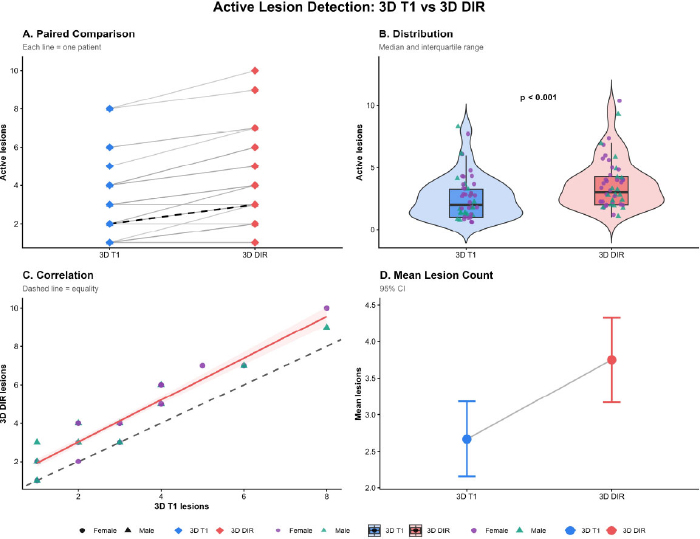
Active lesion detection using 3D T1 and 3D DIR.
(A) Paired lesion counts for each patient. (B) Distribution of lesion counts with individual observations. (C) Correlation between 3D T1 and 3D DIR lesion counts; the dashed line indicates equality and the solid line the fitted regression. (D) Mean lesion counts with 95% CIs. 3D DIR detected significantly more active lesions than 3D T1 (p < 0.001).
Mixed-effects negative binomial regression was performed to evaluate the association between imaging modality and the number of detected lesions while accounting for within-patient correlation (Table 3).
| Variable | IRR | 95% CI | p-value |
|---|---|---|---|
| Imaging modality (3D DIR vs. 3D FLAIR) | 1.55 | 1.20 - 1.99 | 0.001 |
| Age (per year increase) | 1.01 | 0.99 - 1.03 | 0.404 |
| Gender | 1.20 | 0.86 - 1.68 | 0.288 |
3D DIR demonstrated a significantly higher lesion detection rate compared with 3D FLAIR. The incidence rate of detected lesions was approximately 55% higher with 3D DIR (IRR 1.55, 95% CI 1.20-"1.99, p < 0.001).
Neither age nor gender showed a statistically significant association with lesion detection. The likelihood ratio test confirmed significant heterogeneity between patients, supporting the use of a mixed-effects model (p < 0.001).
After adjustment for age and gender, marginal estimates indicated that the mean number of detected lesions was 2.01 (95% CI 1.61-"2.4) for 3D FLAIR and 3.1 (95% CI 2.61-"3.6) for 3D DIR (Table 4).
| Modality | Adjusted Mean | 95% CI | p-value |
|---|---|---|---|
| 3D DIR | 3.10 | 2.61 - 3.60 | <0.001 |
| 3D FLAIR | 2.01 | 1.61 - 2.40 | <0.001 |
4. DISCUSSION
Accurate detection of cortical and juxtacortical lesions is clinically important because these lesions contribute to the diagnosis of multiple sclerosis, reflect disease burden, and may influence disease monitoring and therapeutic decision-making. Advanced MRI techniques such as 3D DIR have been developed to improve visualization of these clinically relevant lesions and have consistently demonstrated superior detection compared with conventional sequences [14-16].
In this study, 3D DIR detected substantially more juxtacortical lesions than 3D FLAIR, identifying approximately 55% more lesions on average. Neither age nor gender showed a significant association with lesion detection. On average, 3D DIR detected about 3.1 lesions per patient compared with about 2.0 lesions using 3D FLAIR, and most patients showed better detection with 3D DIR. Similarly, Elkholy reported that 3D DIR improves lesion visualization compared with FLAIR in several regions, including infratentorial (2.9 vs. 2.25) and periventricular areas (11.84 vs. 11.31), with a marked increase in intracortical lesion detection (7.12 vs. 1.4). 3D DIR demonstrated higher overall sensitivity and was suggested as a potential alternative with minimal additional scan time [17]. Likewise, in a larger cohort, Abdelrahman evaluated 82 patients and confirmed the superiority of 3D DIR over FLAIR in detecting cortical, juxtacortical, periventricular, subcortical, and infratentorial plaques, while no advantage was observed for deep white or gray matter lesions. Notably, cortical lesion burden correlated with disease duration, relapse frequency, and EDSS score, reinforcing its prognostic significance [18].
The diagnostic gain of 3D DIR was even more pronounced in the study by Abidi, where 3D DIR revealed approximately six times more lesions than FLAIR and ten times more than T2-weighted imaging, with markedly improved lesion-to-white-matter contrast. These results strongly supported incorporating 3D DIR into routine MRI protocols [19]. Comparable findings were described by Mostardeiro in a meta-analysis of 3D acquisitions. Across studies, 3D DIR consistently yielded higher counts for cortical (12.4-40.0 vs. 5.25-27.9), intracortical (1.2-8.0 vs. 1.1-3.1), and infratentorial lesions (2.0-12.0 vs. 1.45-8.4), with pooled estimates favoring 3D DIR over FLAIR, particularly in regions critical for disability accumulation [20]. The higher lesion detection observed with 3D DIR is biologically expected because that sequence suppresses signals from the cerebrospinal fluid and the normal white matter, which improves visualization of small cortical and juxtacortical lesions [20, 21].
This study has several important strengths. First, the paired design, in which each patient underwent both imaging modalities, reduced between-subject variability and allowed direct within-patient comparison. Second, the consistency of findings across patients-with most showing higher lesion detection using 3D DIR-strengthens the credibility and clinical relevance of the results. At the same time, several limitations should be acknowledged. The sample size was modest and derived from a single setting, which may limit the generalizability of the findings. Another limitation of this study is that image interpretation was performed by a single experienced radiologist; therefore, inter-observer agreement was not assessed. In addition, other potentially relevant imaging factors, such as lesion subtype, morphology, anatomical distribution, and diagnostic accuracy, and Clinical factors like Expanded Disability Status Scale (EDSS) scores, disease duration, relapse history, and treatment status, were not evaluated. were beyond the scope of this study and should be evaluated in future work. Finally, the study focused on lesion counts rather than diagnostic accuracy or long-term clinical outcomes.
CONCLUSION
In conclusion, with a paired comparison between imaging modalities, 3D DIR demonstrated a significantly higher lesion detection rate than 3D FLAIR, identifying approximately 55% more lesions after adjustment for age and gender. These findings support the added value of 3D DIR for improving lesion visualization and may have important implications for diagnostic practice.
AUTHORS' CONTRIBUTIONS
It is hereby acknowledged that all authors have accepted responsibility for the manuscript's content and consented to its submission. They have meticulously reviewed all results and unanimously approved the final version of the manuscript.
LIST OF ABBREVIATIONS
| DIR | = Double Inversion Recovery |
| MS | = Multiple Sclerosis |
| CNS | = Central Nervous System |
| MRI | = Magnetic Resonance Imaging |
| FLAIR | = Fluid-Attenuated Inversion Recovery |
ETHICS APPROVAL AND CONSENT TO PARTICIPATE
The study received ethical approval from the Ethical Committee of the University of Baghdad, Baghdad, Iraq (Approval No. 168, 2025).
HUMAN AND ANIMAL RIGHTS
All human research procedures followed were in accordance with the ethical standards of the committee responsible for human experimentation (institutional and national), and with the Helsinki Declaration of 1975, as revised in 2013.
AVAILABILITY OF DATA AND MATERIALS
The data supporting the findings of this article are available from the corresponding author [M.T.M] upon reasonable request. The dataset has not been deposited in a public repository.
ACKNOWLEDGEMENTS
Declared none.